
Most Popular
-
1
Blinken calls on China to press N. Korea to end its 'dangerous' behavior

-
2
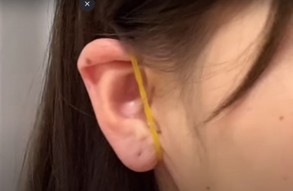
New celebrity-endorsed therapy for face contouring requires only a pair of rubber bands
-
3
Tensions heighten ahead of first president-opposition chief meeting

-
4
[Weekender] How DDP emerged as an icon of Seoul
![[Weekender] How DDP emerged as an icon of Seoul](//res.heraldm.com/phpwas/restmb_idxmake.php?idx=644&simg=/content/image/2024/04/25/20240425050915_0.jpg&u=)
-
5
Seoul to provide housing subsidy to married couples with newborns


![[Weekender] How DDP emerged as an icon of Seoul](http://res.heraldm.com/phpwas/restmb_idxmake.php?idx=644&simg=/content/image/2024/04/25/20240425050915_0.jpg&u=)

-
6
Doctor group's incoming head renews call for govt. to scrap medical school quota hike for dialogue

-
7
Rapper jailed after public street fight with another rapper

-
8
NewJeans pops out ‘Bubble Gum’ video amid troubles at agency

-
9
[Music in drama] An ode to childhood trauma
![[Music in drama] An ode to childhood trauma](//res.heraldm.com/phpwas/restmb_idxmake.php?idx=644&simg=/content/image/2024/04/25/20240425050929_0.jpg&u=)
-
10
Woman gets suspended term for injuring boyfriend with knife


![[Music in drama] An ode to childhood trauma](http://res.heraldm.com/phpwas/restmb_idxmake.php?idx=644&simg=/content/image/2024/04/25/20240425050929_0.jpg&u=)


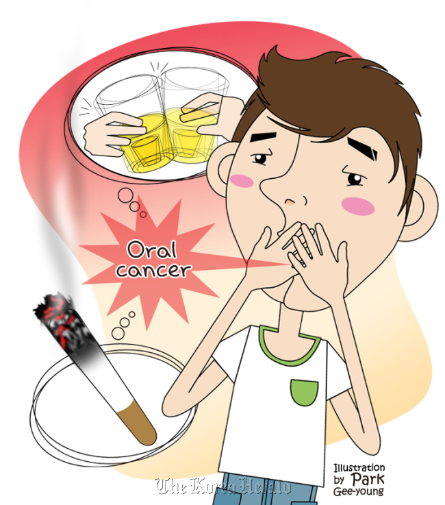
According to the 2008 cancer figures from the National Cancer Center in Korea, there are 2,557 oral cancer cases on average every year, which accounts for 1.4 percent of all cancer patients. The incidence of oral cancer is 5.2 cases per 100,000 persons per year. Oral cancer is one cancer with poor treatment prospects. Since its five-year survival rate of 56 percent in the early 90s, the survival rate has seen little improvement until recently, due to the complicated anatomical location and difficulties in post-surgical reconstruction.
See a specialist regarding unusual lesions in your oral cavity. Do not ignore any painless lumps in your neck.
If symptoms like white bumps (leukoplakia) or red bumps (erythema) are seen on the tongue, the mucous membrane of the oral cavity or of the gingival (gums), this can be a pre-cancerous lesion or an infiltrating carcinoma at its early stage. Also, if there is a firm lump anywhere in the oral cavity or bleeding caused by damaged mucous membrane, this may indicate a growing, late-stage infiltrating carcinoma. In extreme cases, there can be a growing, painless lump without any symptoms and this is often seen in patients if there is metastasis to cervical lymph nodes. As oral cancer progresses, the patients experience difficulties in speaking and ingesting foods, and even in breathing. It also involves discomfort caused by pain and bleeding. Oral cancer is a serious disease where its symptoms can have complex effects and lead to death.
The disease can be diagnosed through a tissue biopsy and its severity assessed by radiological investigation.
The diagnosis is made by taking a biopsy from a suspected lesion in the oral cavity as well as a lump in the neck suspected of lymph node metastasis. Then radiological investigations including CT, MRI and PET scans are performed to assess the severity of the diagnosed oral cancer and the treatment plan is made according to the results.
Early detection and treatment are most important.
It is possible to treat a pre-cancerous lesion (leukoplakia and erythema) detected at a very early stage by removing the localized lesion. In cases of oral cancer detected at a relatively early stage, it is possible to excise the cancer surgically with minimal post-operative disability. However in progressed oral cancer, the excision of the lesion results in severe loss of oral function and it is necessary to perform reconstructive surgery that uses the patient’s muscle, skin and bone to reconstruct the lost structures of the oral cavity after the excision. In addition, the progressed lesion often has not only the localized lesion in the oral cavity but also metastasis to the lymph nodes and therefore, these lymph node metastasis need to be checked before the surgery and removed with the lesion by lymph node excision. Nevertheless, there are cases when the lymph node metastases are not found in the investigations before the surgery and this is believed to be a major cause of recurrence.
Complications with cervical surgery can be reduced by individualized treatments to detect unfound lymph node metastases using sentinel lymph node biopsy; 70 percent of early-stage oral cancer cases are found to have no metastases in the investigations before the surgery.
The sentinel lymph node indicates the first lymph node to be metastasized when cancer cells spread to lymph nodes in an early-stage cancer as well as in breast cancer. The tumor in the oral cavity is injected with a radioactive marker substance and the lymph node to which the injected substance spreads is detected by a radioactive probe. A tissue biopsy is selectively performed and if the biopsy result shows metastases, a widespread cervical lymph node excision is performed accordingly. If not, minimal intervention is used to reduce the surgical area which in turn minimizes the post-operative complications.
The patient may receive surgery, radiotherapy and chemotherapy together.
Based on the progression of the cancer and the surgical findings, radiotherapy and/or chemotherapy may be performed together. In some cases, surgical treatment may also be given in addition to radiotherapy and/or chemotherapy.
Quit smoking. Reduce drinking. Take care of oral hygiene.
Although the exact etiology and mechanism of oral cancer has not been clearly revealed, the biggest carcinogenic factor is smoking. Approximately 90 percent of cases of oral cancer are related to smoking. Smokers, compared to non-smokers, have a 2-4 times higher risk of getting oral cancer. The risk is proportional to the amount and the period of smoking. When exposed to smoking, a gradual morphological change happens in the mucous membrane of the oral cavity over a long period of time, and this causes cancer. This morphological change is reversible; therefore quitting smoking reduces the risk of cancer development. Drinking is also a carcinogenic factor. People who smoke and drink excessively have both risk factors. This promotes the development of oral cancer and the person who does both has a 6-15 times higher risk of contracting oral cancer compared to a person who does not smoke or drink. In addition, vitamin deficiency, iron deficiency anemia, ultraviolet rays, poor oral and dental hygiene are related to the development of oral cancer.
Oral cancer is a dangerous disease. It has a relatively low survival rate and it results in serious disabilities after treatment. It also has a serious effect on the quality of life due to aesthetic issues. This is why early detection and prevention are the most important. Even in a progressed lesion, if the treatment and rehabilitation is performed under precise plan, it is believed that the cancer can be overcome.
By Baek Chung-hwan
The author is a professor of Otorhinolaryngology- Head&Neck Surgery at Sungkyunkwan University School of Medicine and doctor at Samsung Medical Center ― Ed.
See a specialist regarding unusual lesions in your oral cavity. Do not ignore any painless lumps in your neck.
If symptoms like white bumps (leukoplakia) or red bumps (erythema) are seen on the tongue, the mucous membrane of the oral cavity or of the gingival (gums), this can be a pre-cancerous lesion or an infiltrating carcinoma at its early stage. Also, if there is a firm lump anywhere in the oral cavity or bleeding caused by damaged mucous membrane, this may indicate a growing, late-stage infiltrating carcinoma. In extreme cases, there can be a growing, painless lump without any symptoms and this is often seen in patients if there is metastasis to cervical lymph nodes. As oral cancer progresses, the patients experience difficulties in speaking and ingesting foods, and even in breathing. It also involves discomfort caused by pain and bleeding. Oral cancer is a serious disease where its symptoms can have complex effects and lead to death.

The disease can be diagnosed through a tissue biopsy and its severity assessed by radiological investigation.
The diagnosis is made by taking a biopsy from a suspected lesion in the oral cavity as well as a lump in the neck suspected of lymph node metastasis. Then radiological investigations including CT, MRI and PET scans are performed to assess the severity of the diagnosed oral cancer and the treatment plan is made according to the results.
Early detection and treatment are most important.
It is possible to treat a pre-cancerous lesion (leukoplakia and erythema) detected at a very early stage by removing the localized lesion. In cases of oral cancer detected at a relatively early stage, it is possible to excise the cancer surgically with minimal post-operative disability. However in progressed oral cancer, the excision of the lesion results in severe loss of oral function and it is necessary to perform reconstructive surgery that uses the patient’s muscle, skin and bone to reconstruct the lost structures of the oral cavity after the excision. In addition, the progressed lesion often has not only the localized lesion in the oral cavity but also metastasis to the lymph nodes and therefore, these lymph node metastasis need to be checked before the surgery and removed with the lesion by lymph node excision. Nevertheless, there are cases when the lymph node metastases are not found in the investigations before the surgery and this is believed to be a major cause of recurrence.
Complications with cervical surgery can be reduced by individualized treatments to detect unfound lymph node metastases using sentinel lymph node biopsy; 70 percent of early-stage oral cancer cases are found to have no metastases in the investigations before the surgery.
The sentinel lymph node indicates the first lymph node to be metastasized when cancer cells spread to lymph nodes in an early-stage cancer as well as in breast cancer. The tumor in the oral cavity is injected with a radioactive marker substance and the lymph node to which the injected substance spreads is detected by a radioactive probe. A tissue biopsy is selectively performed and if the biopsy result shows metastases, a widespread cervical lymph node excision is performed accordingly. If not, minimal intervention is used to reduce the surgical area which in turn minimizes the post-operative complications.
The patient may receive surgery, radiotherapy and chemotherapy together.
Based on the progression of the cancer and the surgical findings, radiotherapy and/or chemotherapy may be performed together. In some cases, surgical treatment may also be given in addition to radiotherapy and/or chemotherapy.
Quit smoking. Reduce drinking. Take care of oral hygiene.
Although the exact etiology and mechanism of oral cancer has not been clearly revealed, the biggest carcinogenic factor is smoking. Approximately 90 percent of cases of oral cancer are related to smoking. Smokers, compared to non-smokers, have a 2-4 times higher risk of getting oral cancer. The risk is proportional to the amount and the period of smoking. When exposed to smoking, a gradual morphological change happens in the mucous membrane of the oral cavity over a long period of time, and this causes cancer. This morphological change is reversible; therefore quitting smoking reduces the risk of cancer development. Drinking is also a carcinogenic factor. People who smoke and drink excessively have both risk factors. This promotes the development of oral cancer and the person who does both has a 6-15 times higher risk of contracting oral cancer compared to a person who does not smoke or drink. In addition, vitamin deficiency, iron deficiency anemia, ultraviolet rays, poor oral and dental hygiene are related to the development of oral cancer.
Oral cancer is a dangerous disease. It has a relatively low survival rate and it results in serious disabilities after treatment. It also has a serious effect on the quality of life due to aesthetic issues. This is why early detection and prevention are the most important. Even in a progressed lesion, if the treatment and rehabilitation is performed under precise plan, it is believed that the cancer can be overcome.
By Baek Chung-hwan
The author is a professor of Otorhinolaryngology- Head&Neck Surgery at Sungkyunkwan University School of Medicine and doctor at Samsung Medical Center ― Ed.
-
Articles by Korea Herald


![[Herald Interview] Mistakes turn into blessings in street performance, director says](http://res.heraldm.com/phpwas/restmb_idxmake.php?idx=652&simg=/content/image/2024/04/28/20240428050150_0.jpg&u=20240428174656)