
Most Popular


-
6
Actor’s excessive airport security sparks probe into human rights violations

-
7
Man who let his father die due to financial difficulties to be released on parole

-
8
Actor Yoo Ah-in accused of sexual attack

-
9
S. Korea, China shifting from tensions to cooperation: Seoul

-
10
LG Electronics achieves record earnings in Q2


[From the Scene] Digitized hospital provides glimpse into next-generation diagnoses
Singapore General Hospital, housing one of world’s largest installation for digital pathology systems, stresses the need to advance health care tech, digitize data for diagnostic agility and accuracy
By Lee Yoon-seoPublished : July 27, 2023 - 12:01
By Lee Yoon-seo
Korea Herald correspondent
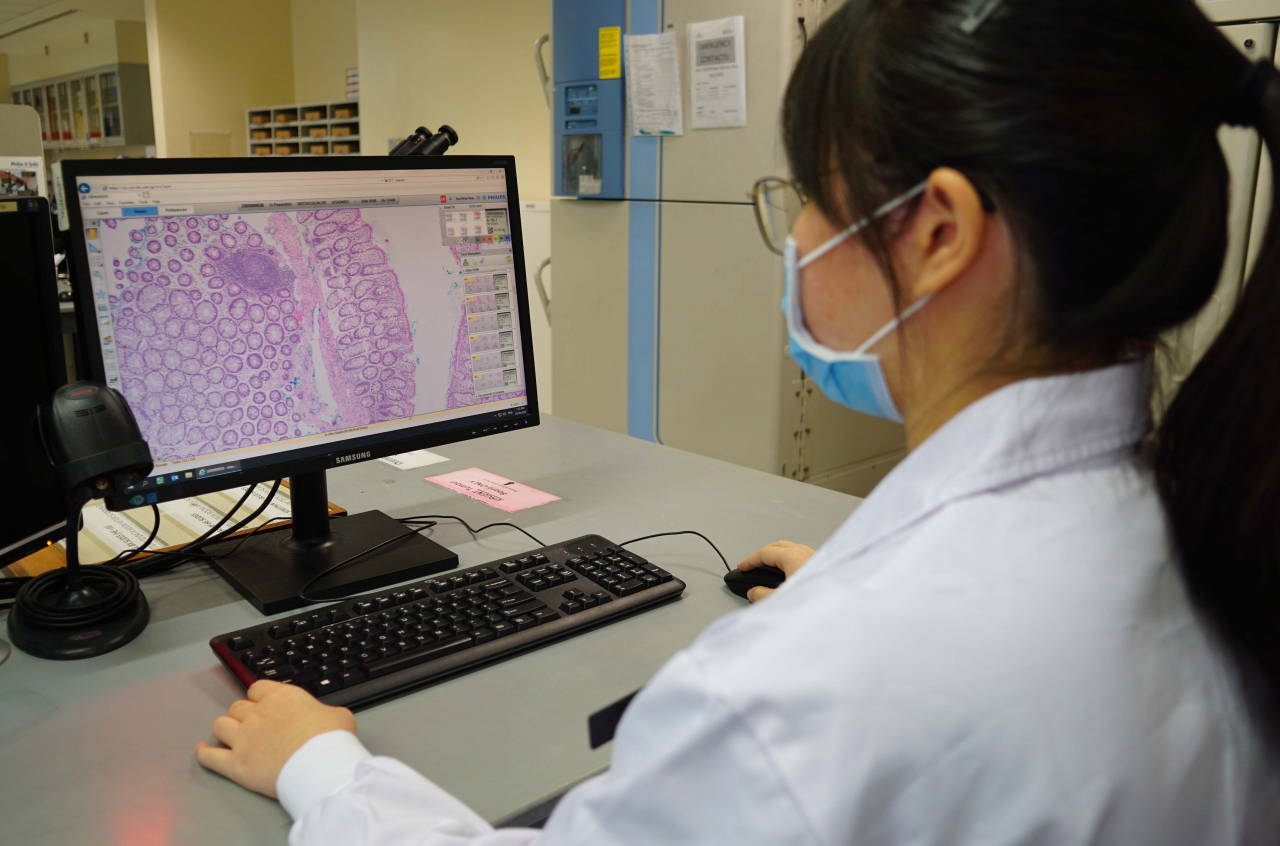
SINGAPORE -- A masked resident pores over a piece of uterine fibroid -- a tumor developed in the uterus -- and cautiously slices it with a scalpel.
The thinly sectioned tissue specimen, after going through various processes including chemical treatment, flattening and staining, is then put on a small glass slide by a scientist.
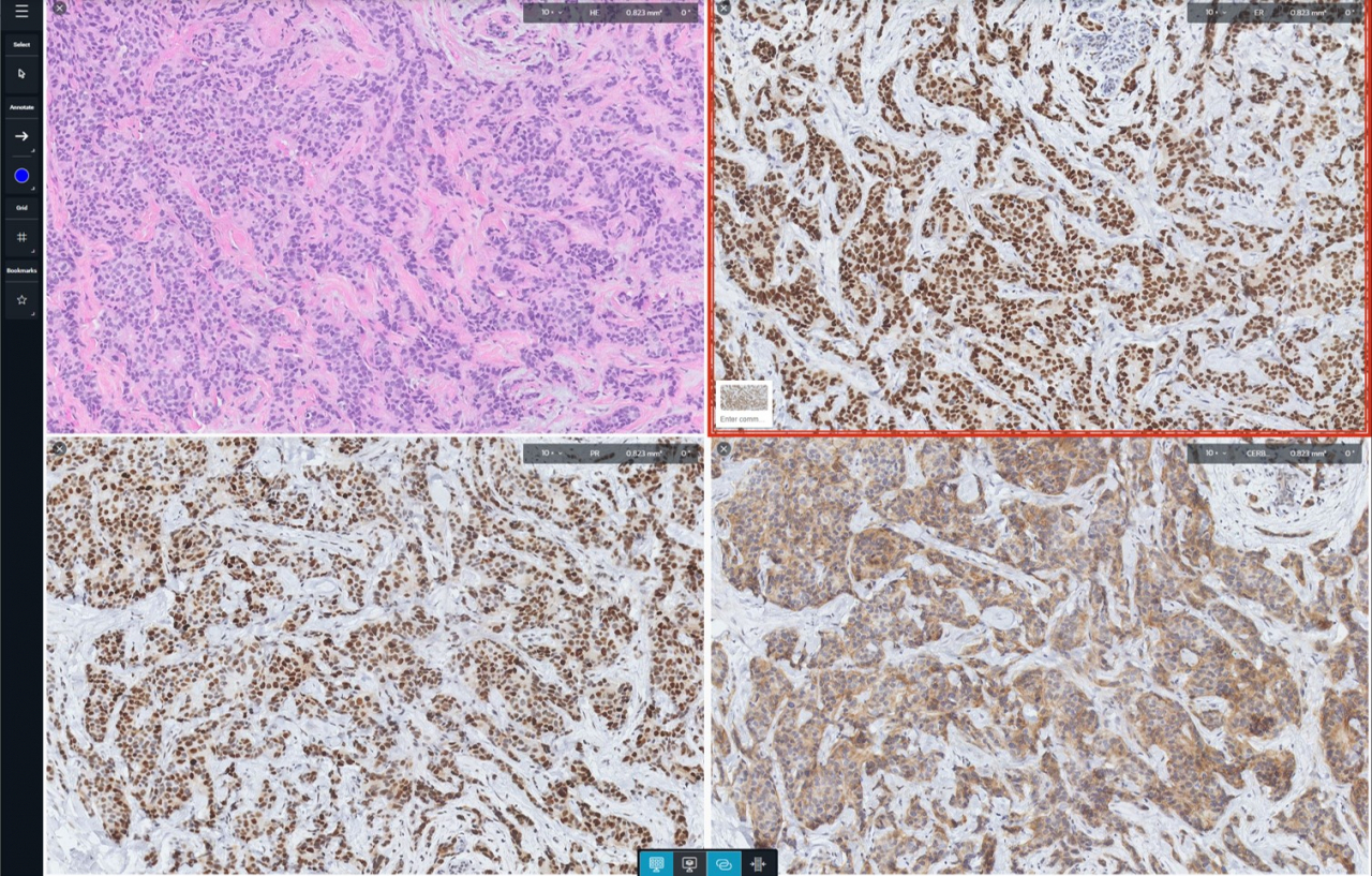
With whole slide imaging, the tissue on the glass slide is virtually copied into a highly magnified image, to be uploaded onto a digital system.
The image is then shared across a slew of pathologists, to be examined in depth to determine if the cells in the tissue show abnormal signs of cell activity that could connote contraction of uterine cancer.
According to Cheng Chee Leong, the head and senior consultant anatomical pathology at Singapore General Hospital, such digitization of tissue analysis is breaking through the constraints pathologists currently suffer from when trying to perform tissue analysis -- a critical part of making a slew of diagnoses for diseases, genetic research, and even a development of treatment protocols. According to Leong, currently, SGH stands as one of the hospitals in the world with the largest installation of whole slide imaging processes.
"For the past century, the use of glass and microscopes have been called to the practice of pathology and it is still the current standard (for tissue analysis)," said Leong.
However, examining tissues on glass slides present an array of complications for pathologists across the world, according to Leong.
Tissues on glass slides fade if they are left there for too long, which poses a risk of losing precious samples for diagnosis, he said.
"Furthermore, the physical constraint of the glass slides was such that we can only use glass slides for one of the many purposes. We use (tissue images) for consultation, diagnosis, teaching, research and so on. However, the glass slides could be only used for one purpose at a time," he added.
"And when you were not using the glass slides, they were stored somewhere far away, sometimes off-site. They were not readily retrievable. So, sometimes it could actually take days or weeks before you could obtain the glass slides. When you wanted to refer to a previous diagnosis, (such a procedure) was very inconvenient."
However, with the help of advancements in the health care technology industry, Leong said such issues are handily solved as digitized images of tissues allow for complete preservation of materials, increased accessibility to data and multipurpose use of one tissue specimen.
"Whole slide images also allow multiple pathologists to access the same material at the same time, even if we are apart. Before, we needed to physically bring a glass slide to discuss the matter with fellow colleagues," Leong added.
"Now, pathologists can even share cases internationally, allowing collaboration in diagnosis teaching and research."
He stressed that such state-of-the-art technology most importantly allows pathologists to deliver the slides more securely and accurately, increasing the chances of giving patients an accurate diagnosis in a timely manner.
Leong said the digitization of a single procedure paved the way for the rise of various advanced digitized mechanisms.
Currently, Leong said SGH is working to further unlock the digitalized slides' potential with computational technology.
Work is in progress to streamline the process of machines performing automatic diagnoses with retrieval of known diagnoses of diseases, based on databases of whole slide images.
In addition, for digitized breast tissue images, projects are in place in to have machines learn algorithms that can automatically differentiate benign breast tumors from those that tend to grow more rapidly and have a chance of being malignant.
"Because we are able to work with the digital images, (we are able to work) on projects like artificial intelligence, modeling and recognition of certain disease activities (by using whole slide imaging)," he said, stressing that the adoption of digital technology in medical processes were essential for providing more accurate and timely diagnoses.


![[Today’s K-pop] Treasure to publish magazine for debut anniversary](http://res.heraldm.com/phpwas/restmb_idxmake.php?idx=642&simg=/content/image/2024/07/26/20240726050551_0.jpg&u=)